See What Is Under the Surface
Imaging and analysis that shows what your skin is doing beneath what you can see, so the plan is built on data rather than impression.
A Measurement, Not a Sales Tool
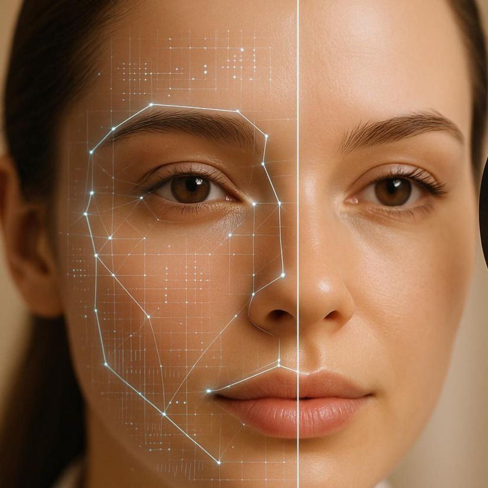
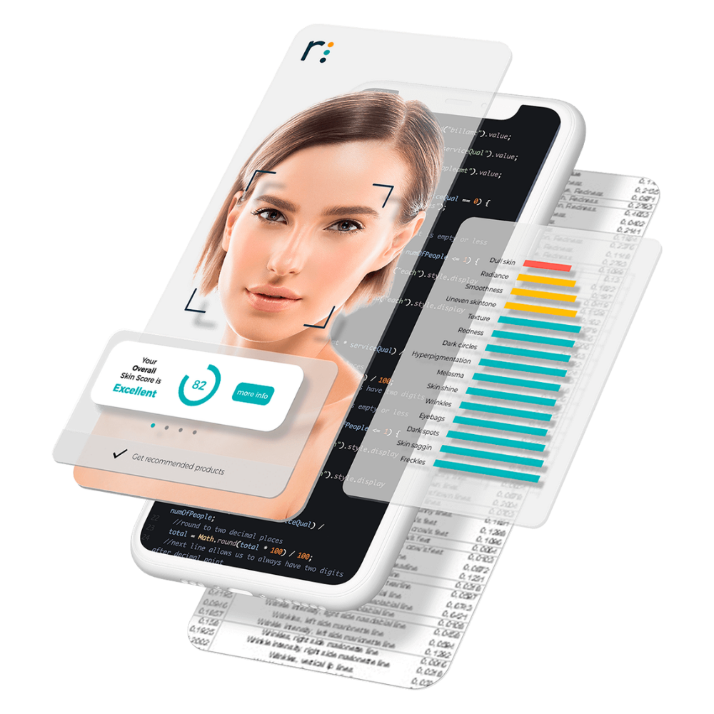
Skin analysis imaging captures your skin under controlled lighting and processes it to surface things the naked eye cannot reliably judge: pigment sitting below the surface, vascular patterns, pore distribution, texture and the early signs of photodamage.
The value is that it replaces impression with information. Two people can look at the same face and disagree about whether pigment or texture is the dominant problem. An image analyzed consistently removes that argument, and it removes it in both directions.
It is also the fairest way to judge whether treatment is working. Skin changes slowly, memory is unreliable, and a mirror does not keep records. Imaging you at the start and again later tells you the truth about whether the plan is earning its place, including when the answer is that it is not.
A Good Fit If Any of This Sounds Familiar
This is an assessment tool. We use it to build a plan and to check it, not to generate a shopping list.
Three Steps, Start to Finish
We Capture the Images
A short session under controlled lighting. Nothing touches your skin and there is nothing to recover from.
We Review It Together
On screen, with you, explaining what each layer is showing and what it does and does not mean.
The Plan Follows the Findings
Including the parts where the finding is that you do not need much. That happens more often than people expect.
Sometimes the Data Says You Do Not Need Us
An assessment that can only ever conclude you need treatment is not an assessment. It is a sales process with a camera.
Twenty Minutes and a Clear Picture
What the Imaging Can and Cannot Tell You
Different wavelengths of light penetrate skin to different depths and are absorbed differently by melanin and hemoglobin. Capturing under controlled conditions allows pigment and vascular structures below the visible surface to be separated and mapped, which is why an image can show damage a mirror cannot.
Analysis then compares what was captured against reference data to characterise things like pore distribution, surface texture and pigment density. It is a consistent way of describing skin rather than a diagnosis.
It does not diagnose disease. Anything clinically suspicious, including changing or irregular lesions, needs proper medical evaluation rather than a skin analysis, and we will refer or examine appropriately rather than let an imaging session stand in for that.
What People Ask Us First
No. It characterises skin and gives us a consistent baseline. Anything that looks clinically concerning is handled as a medical matter, not as an imaging finding.
Come in without makeup if you can. Beyond that, nothing.
We use it to build a plan, and a plan that says wait or do less is a legitimate outcome. If the imaging does not support treating something, we will not recommend treating it.
After a meaningful course of treatment, or annually if you are tracking prevention. Repeating it too often shows noise rather than change.
It can show damage already present that has not surfaced yet, which is useful and is not the same as prediction. We will be careful about that distinction rather than overstate it.
Often Considered Alongside This
Start With What Your Skin Is Actually Doing.
Twenty minutes of assessment before you spend anything is the cheapest decision on this entire site.
4211 N. Buffalo Road, Suite 15, Orchard Park, NY 14127
